Equipments
Diagnostics Equipments
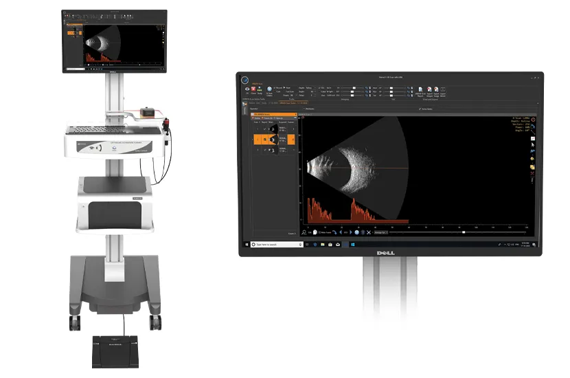
Appasamy Marvel - Ocular Ultrasound
Ophthalmic ultrasonography (Ocular USG) is sometimes needed when the retina or posterior segment is not visible by the conventional examination techniques. Anjani Eye Hospital acquired the Appasamy Marvel Ocular USG which is the best of the range USG machines, in 2020. Ocular USG may sometimes be needed to diagnose some retinal pathologies like Retinoblastoma and other intra / extra ocular tumors, intraocular foreign bodies etc.
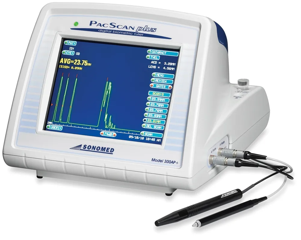
Sonomed Pacscan - A scan Biometer
IOL power calculation is the most crucial part of pre operative evaluations for cataract surgery to give patient’s the best visual outcome. Anjani Eye hospital has been using A-scan biometry for its patient’s since 1984-85 and has been upgrading its instruments to give the best results.
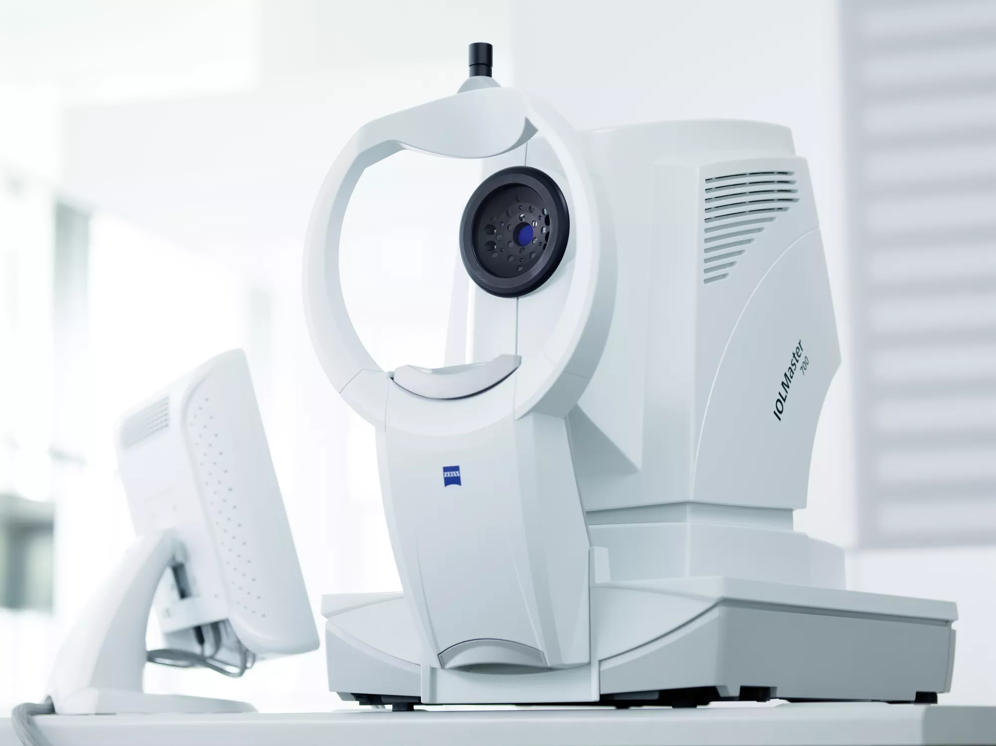
Zeiss IOL Master 700 - Optical Biometer
To Improve the cataract surgery outcomes for the patient’s visiting and getting treated at Anjani Eye Hospital, we upgraded to Zeiss IOL Master 700 from its predecessor. The optical bio-meter uses very low intensity non harmful laser beams to acquire various measurements and in turn accurately calculate IOL power required for that eye.
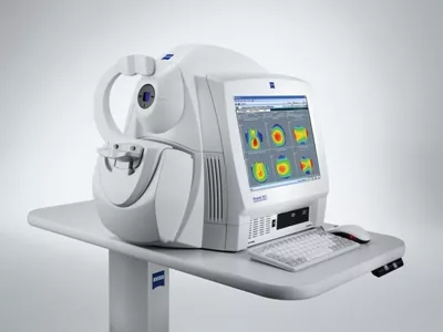
Zeiss Visante OCT with the Visante OMNI - Anterior segment OCT
The Visante OCT uses a non-contact technique to provide sharp, highly detailed images and precise biometrics of the anterior segment, including corneal shape and angle information — without the need for ocular anesthesia or time-consuming water baths. Integrating proven anterior topography from the ATLAS Corneal Topographer with precision OCT pachymetry, Visante omni provides comprehensive anterior and posterior topography with pachymetry analysis for improved patient selection and care.
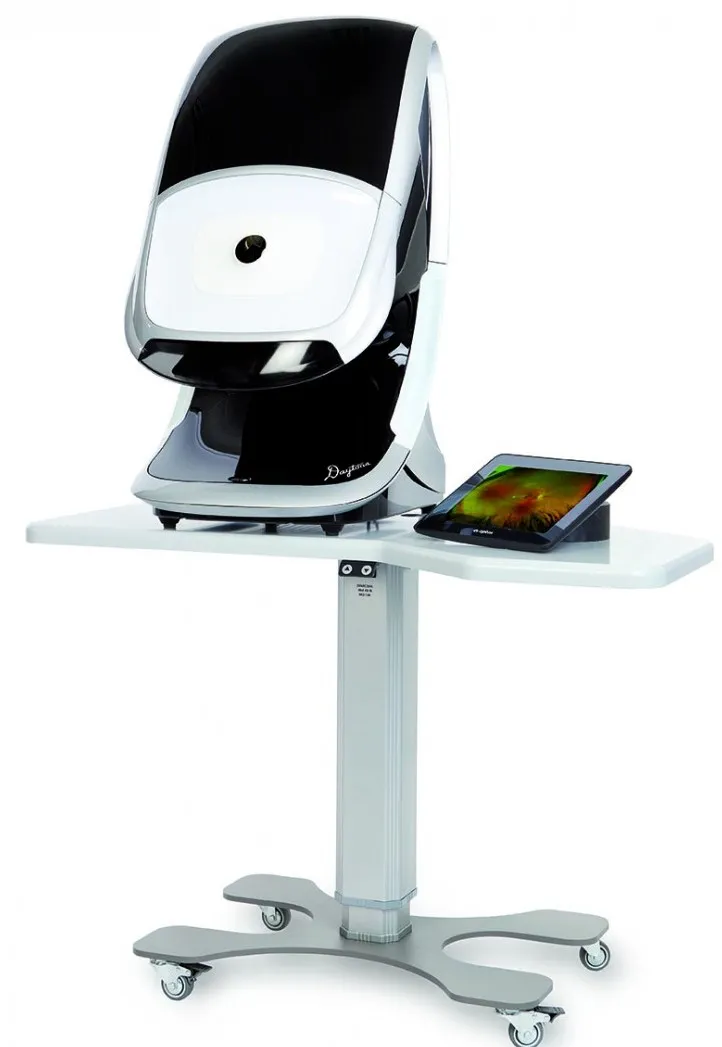
Optos Daytona – Ultra Widefield Retinal Imaging
Widefield retinal imaging by Nikon-Optos produces a 200° single-capture optomap retinal image of unrivaled clarity in less than ½ a second. This fast, easy, patient-friendly, ultra-widefield imaging technology designed for healthy eye screening. To improve practice flow and patient management, Anjani Eye Hospital recently acquired the Optos Daytona in December 2023.
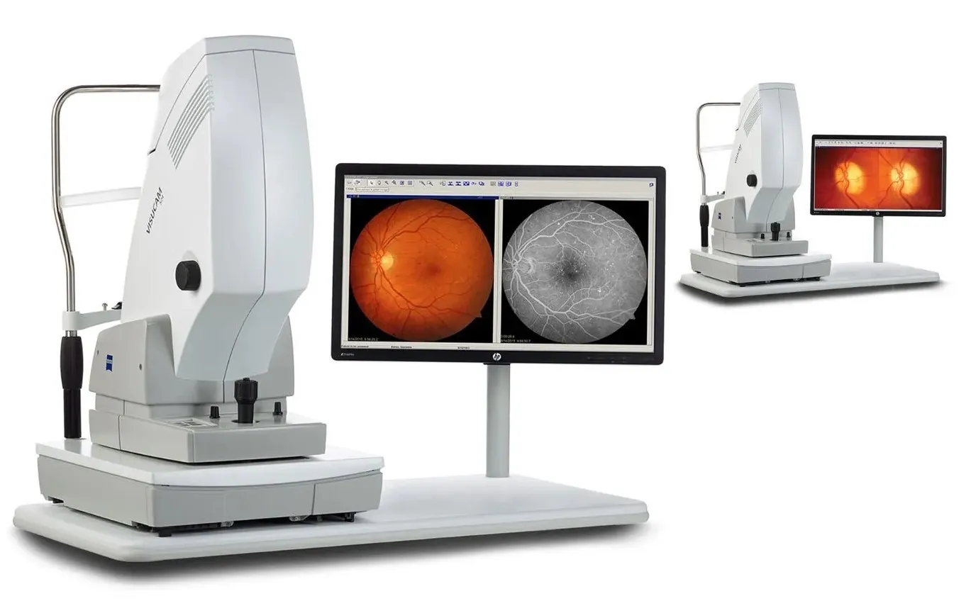
Zeiss Visucam 524 - Fundus Camera
The Visucam 524 Fundus Camera from Zeiss at Anjani Eye Hospital produces brilliant, detail rich images of the retina to effectively diagnose and monitor a broad range of eye conditions – glaucoma, diabetic retinopathy, age related macular degeneration (ARMD). It also has the capability of doing Fundus Fluorescein Angiography (FFA) and Indocyanine Green Angiography (ICGA).
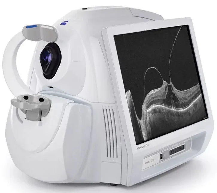
Zeiss Cirrus OCT Angioplex
The Zeiss Cirrus OCT Angioplex at Anjani Eye Hospital includes the latest in retinal and glaucoma diagnostics, with optical coherence tomography (OCT). In addition to the conventional OCT, the cirrus has the capability of OCT angiography (non-invasive OCT angiography (OCTA) technology, to visualize retinal microvasculature and monitor change over time), anterior segment OCT and en face imaging all with the efficiency and reliability.
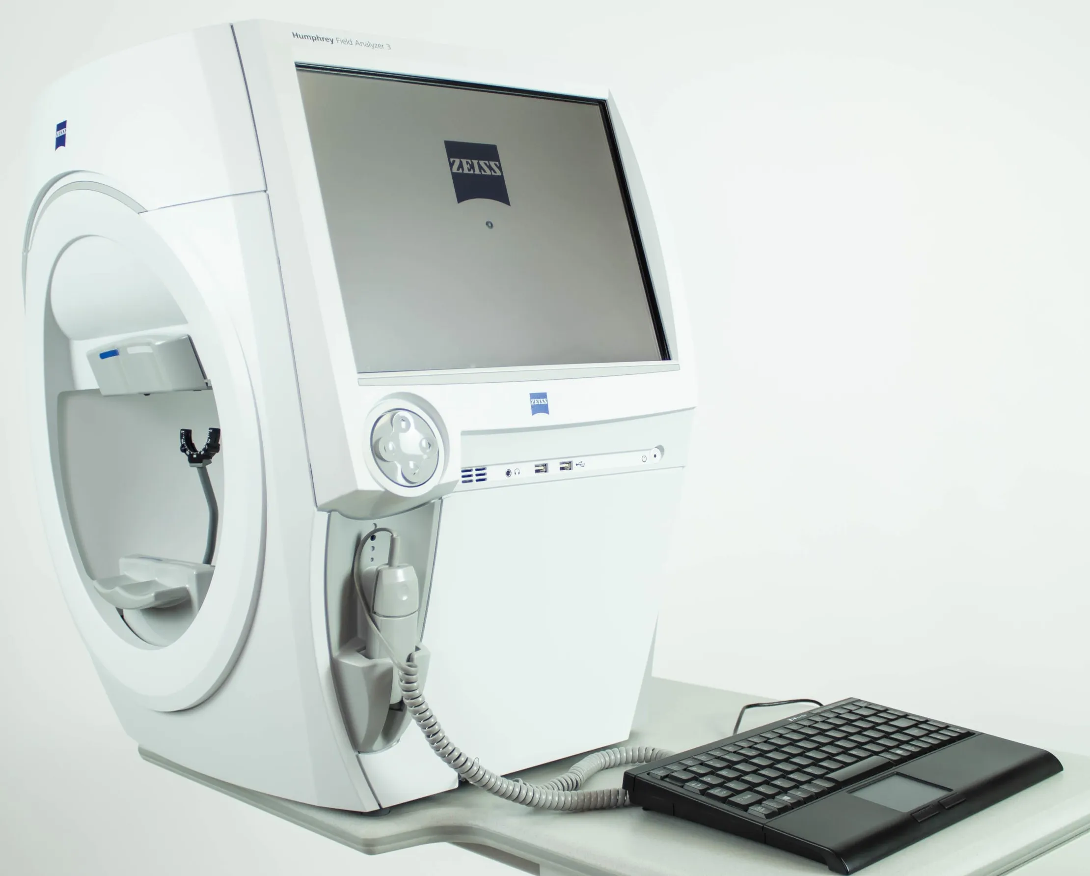
Zeiss Humphrey’s Field Analyzer
Glaucoma diagnostics at Anjani Eye Hospital has always been uncompromised with the use of the gold standard in Perimetry (visual field analysis) with the Humphrey’s Field Analyzer by Zeiss. Not only does it help in diagnosing field defects in glaucoma and neurology, but it also helps in monitoring disease progression.
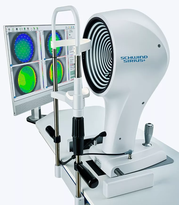
Schwind Sirius+ Corneal Topographer
The Sirius+ corneal topographer by Schwind at Anjani Eye Hospital uses a combination of a sophisticated 3D Scheimpflug camera and well-established Placido topography. The 3D Scheimpflug camera generates pachymetry map of the eye and provides information about corneal thickness. The high-resolution Placido topography allows for a particularly detailed overview of the cornea including corneal aberrations. This practically error free information is used for LASIK, and to diagnose and monitor corneal ectasia (keratoconus).
Lasers Equipments
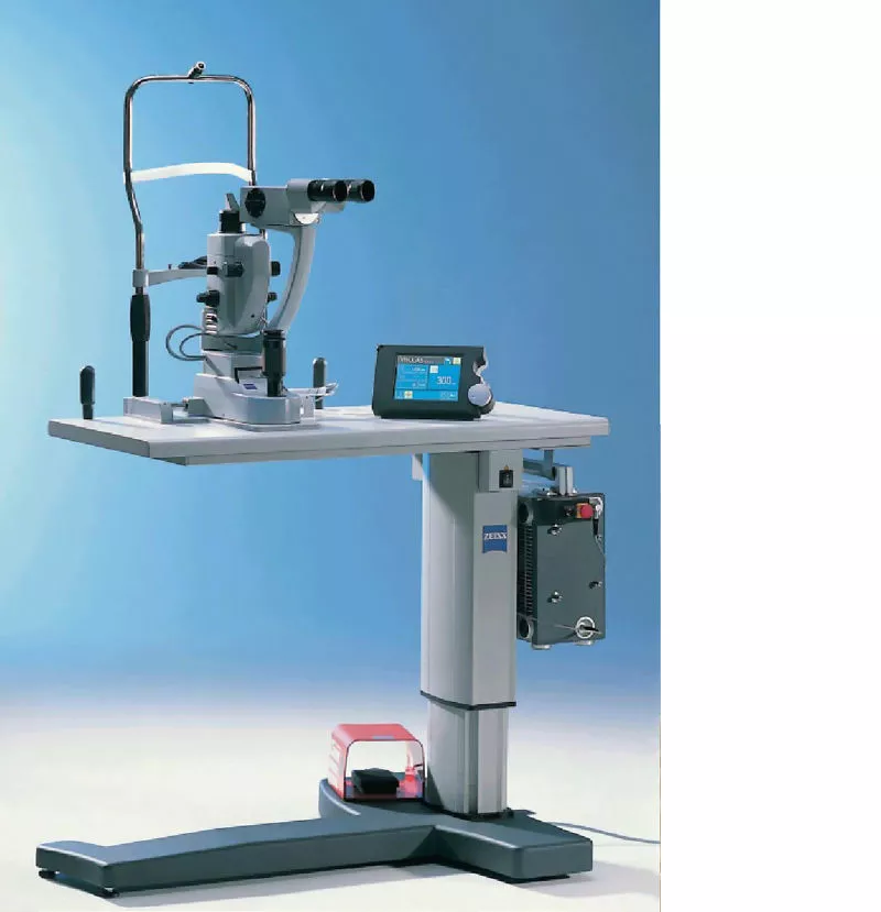
Zeiss VISULAS 532s - Retinal Laser System
Anjani Eye Hospital in its endeavour in delivering the best and highest quality treatment to its patient’s never compromised in the equipment it needed for the same. The Zeiss VISULAS 532s laser system is one such system that is used for Retinal photocoagulation as a treatment of choice in majority of retinal problems. Laser Trabeculoplasty or iridotomy which is a treatment of choice for uncontrolled glaucoma can also be done by this machine.
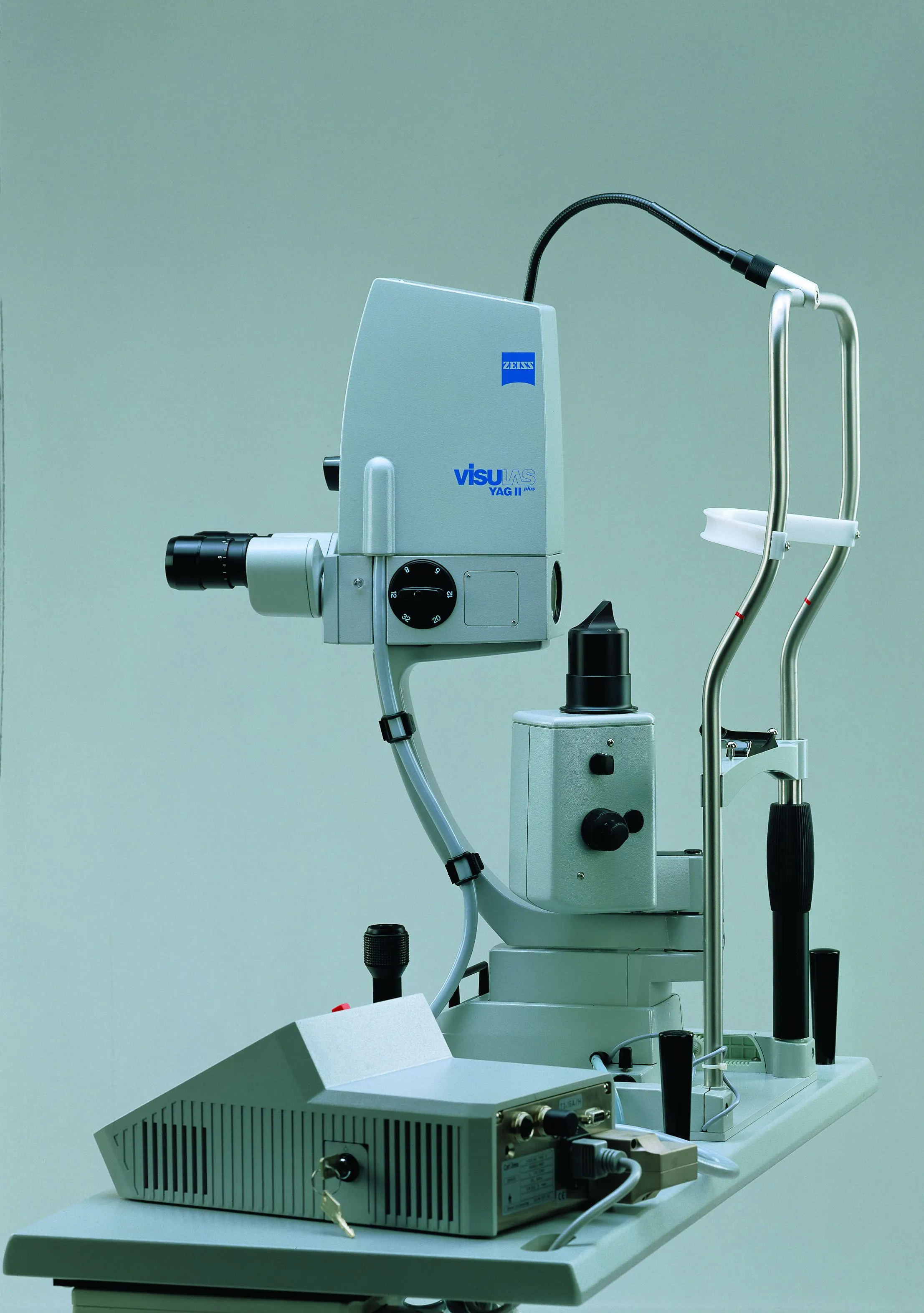
Zeiss VISULAS Nd - YAG Laser system
Most common cause of blurring of vision after a few months of cataract surgery is because of a condition called as Posterior Capsular Opacification (formation of a thin layer behind the implanted lens). NdYAG laser is used for clearing this layer behind the IOL gives instant improvement of vision to these patients. Anjani Eye Hospital acquired the Zeiss VISULAS Nd-YAG laser system for the benefit of its patients. The same laser system can also be used for Peripheral iridotomy to treat certain types of glaucoma.
OPD Equipments
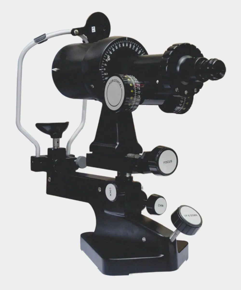
Keratometer
A diagnostic instrument commonly used to measure the curvature of cornea of the eye in the Out Patient Department. The curvature of the cornea is essential in calculating the Lens power of patient’s posted for cataract surgery. It also helps to assess the extent and axis of cylinderical power in patient’s with astigmatism. It also helps in diagnosis of corneal ectatic conditions like Keratoconus, Pellucid Marginal Degeneration etc.
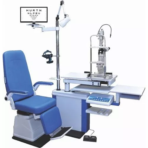
Ophthalmic Chair Unit with Indirect Ophthalmoscope and Slit-lamp Biomicroscope
The Chair unit is an essential equipment needed for routine eye examination in the out patient department. It has a motorised chair which can be moved up, down and reclined (so as to adjust the patient’s height and position during the eye check-up. Indirect Ophthalmoscope helps the ophthalmologist / vireo-retinal surgeon to precisely diagnose retinal disorders. It is especially useful in high myopia, diabetic retinopathy, retinal detachment etc. Slit-lamp Biomicroscope is an essential instrument and is a specialised microscope used in the OPD for routine eye examination. It gives magnified view of the patient’s eye and its various structures and helps the eye doctor in diagnosing and monitoring various eye conditions.
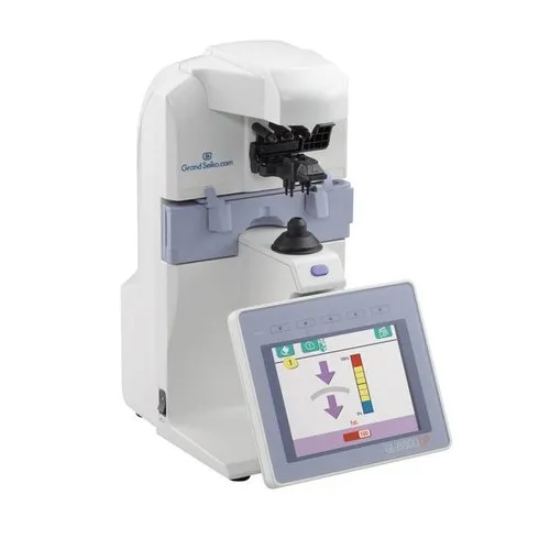
Grand Seiko Auto Lensometer
An Auto-lensometer helps the optometrist in correctly measuring the power of the lens fitted in the spectacle being used by the patient.
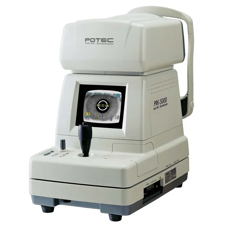
Potec Auto Refractometer
Auto-refractometer helps the optometrist in correctly measuring the power of the glasses needed for the patient’s eye. The optometrist then uses this value for the spectacle correction on the patient’s eye. This instrument also helps in measuring the corneal curvature like the keratometer.
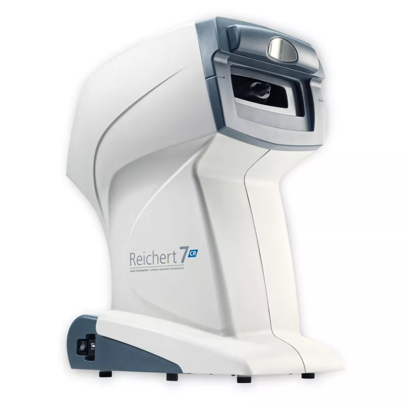
Reichert Non contact tonometer
A Non – Contact Tonometer measures Intra Ocular Pressure (IOP) without touching the eye and is a screening tool used to help in diagnosis Glaucoma.
Surgical Equipments
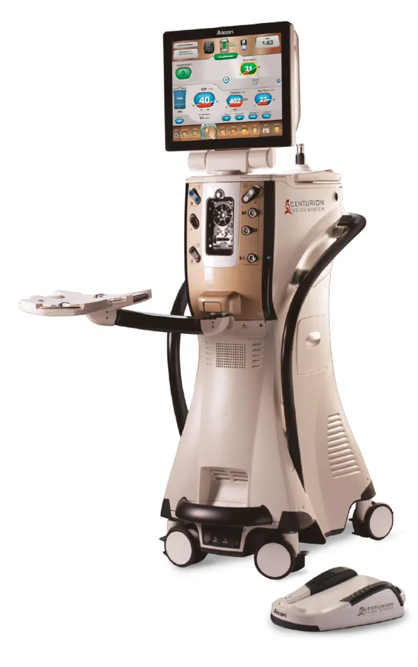
Alcon Centurion Vision System - Phacoemulsification system
Anjani Eye Hospital added this top of the class Phacoemulsification system in 2016 to reduce the risks of complications in cataract surgery and give a better outcome to the patient. It is used during cataract surgery to disintegrate natural lens into smaller particles and to suck the lens matter through a very small incision [Micro Incision Cataract Surgery (MICS)].
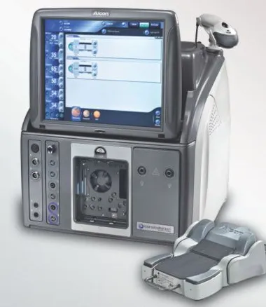
Alcon Constellation Vision system - Vitrectomy system
This is recognised as world’s best operating system for Micro Incision Vitrectomy Surgery (MIVS) [23G, 25G or 27G] and Anjani Eye Hospital has been a proud owner of this system since 2016. This system is used to do Vitreo-retinal surgeries including surgery for retinal detachment, macular hole, surgeries for diabetic retinopathy etc., which are considered as vision saving surgeries.
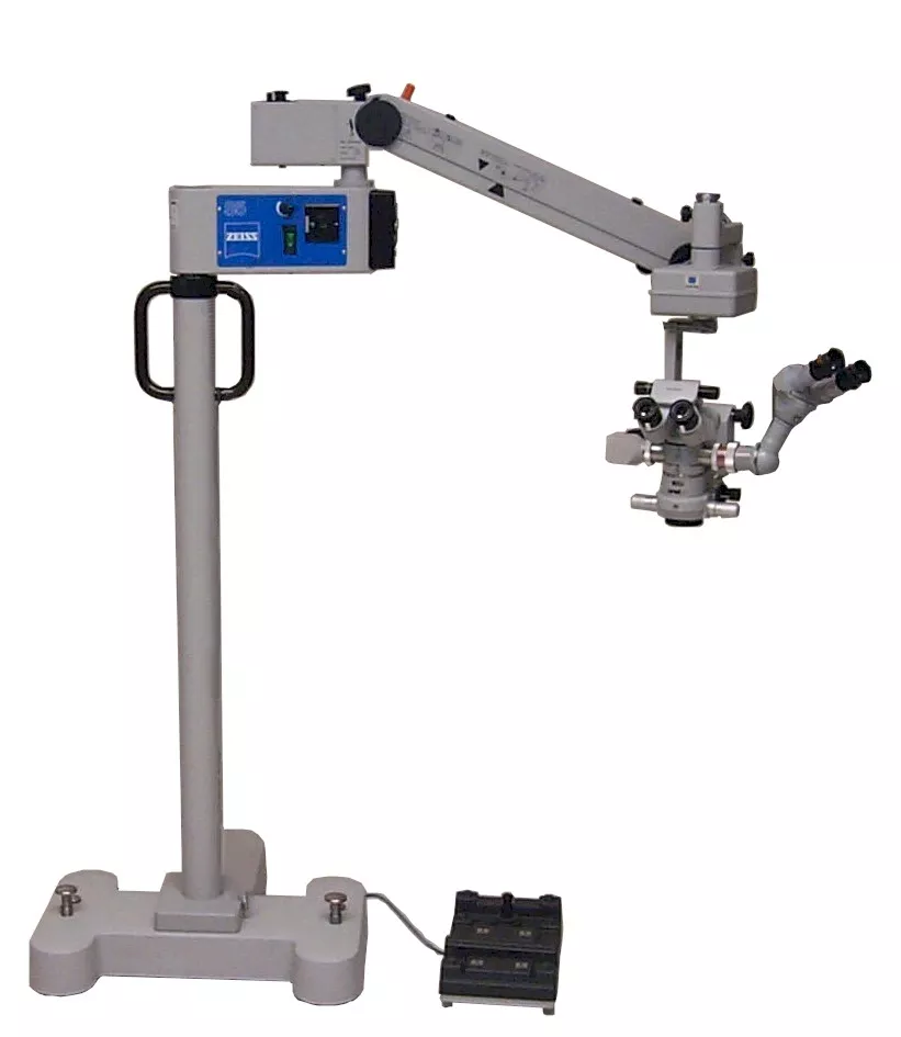
Zeiss Lumera i and Zeiss MDO Operating Microscope
Any eye surgery is done using microscopes. Anjani Eye Hospital in its endeavour to serve its patients with the best quality services has been using Zeiss microscopes with the world class Zeiss optics to give give the operating surgeons very good quality depth of focus. With the surgeons comfortable, the surgical procedure becomes very safe and effective for the patients.
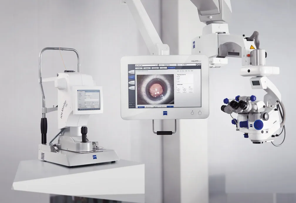
Zeiss Calisto Eye
This advanced system from Zeiss was acquired by Anjani Eye hospital in 2019. It helps surgeons to mark the incision during cataract surgeries especially when Toric IOLS (used for patients of > 1 cylinder astigmatic power) are used. Toric IOLs and location of incision enable surgeons to improve the surgical outcome for the patients in giving crystal clear vision even for high cylindrical power.
Alcon NGENUITY 3D Visualization System
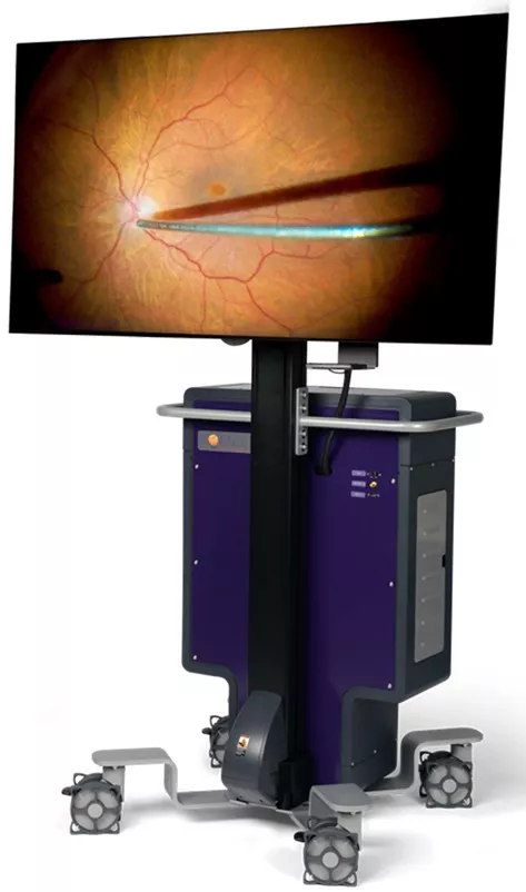
The NGENUITY® 3D Visualization System by ALCON, at Anjani Eye Hospital, is a platform for digitally assisted ophthalmic / eye surgery. It is an adjunct to the surgical microscope, but rather than looking through the microscope eye-piece, the surgeon sees what he or she is doing in 3D on a 55-inch high-definition display. The NGENUITY 3D Visualization System delivers an extended depth of field, up to a 48% increase in magnification, with an edge-to-edge image clarity. It’s digital image processing allows to operate under low lighting condition using only the amount of light needed, which results in reduced patient risk factor.
Contact
Address:20, Farmland, Central Bazar Road, Near Lokmat Square, New Ramdaspeth, Nagpur – 440010, Maharashtra, India. Phone:+91 712 2425 839/2425 869/2425 899 For Appointment:+91 78755 20005 WhatsApp: +91 78755 10002 Email:anjanieyehospital1976@gmail.com Hospital Working Hours:7:30 AM – 5:30 PM (Monday to Friday) 7:30 AM – 3:30 PM (Saturday) Sunday Closed
Copyright © 2025. All rights reserved. Built bySHOUT IN & OUT
Privacy Policy
