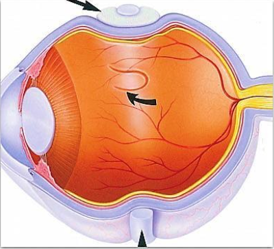
Retina is the light sensitive layer of tissue that lines the inside of the eye and sends visual messages through the optic nerve to the brain. Retinal Detachment is a disorder of the eye in which the retina separates from the underlying layer. If not treated promptly, retinal detachment can cause permanent vision loss. In about 7% of cases both eyes are affected.
What are the types of Retinal Detachment?
There are 3 types of Retinal Detachment:
Rhegmatogenous Retinal Detachment – This occurs due to a break in the retina called retinal tear that allows fluid to pass from the vitreous into the space beneath the retina.
Exudative / Serous / Secondary Retinal Detachment – This occurs due to inflammation / injury / vascular abnormalities leading to accumulation of fluid beneath the retina without the presence of retinal break.
Tractional Retinal Detachment – This occurs when fibrous / fibro-vascular tissue caused by an injury / inflammation / neo-vascularization pulls the retina towards itself.
What are the risk factors for Retinal Detachment?
The common risk factors for Retinal Detachment include – high myopia, trauma, and complications from eye surgery or family history. Other risk factors include – diabetic retinopathy, eclampsia, malignant hypertension, eye cancer etc… (These risk factors usually lead to exudative retinal detachment or Tractional retinal detachment.)
What are the signs and symptoms of Retinal Detachment?
Symptoms include a sudden increase in the number of floaters, flashes of light and worsening of the outer part of the side vision (commonly described as a curtain falling over part of the vision).
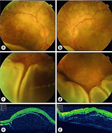
How is Retinal Detachment diagnosed?
Retinal Detachment can be easily diagnosed by an ophthalmologist on ophthalmoscopy. However, the following tests are done by an ophthalmologist when you present with sudden decrease in your vision.
Incase your eye doctor is unable to visualize your fundus due to cataract or vitreous hemorrhage, he/she may advice for a ultrasound (USG B-Scan) of your eye to confirm the diagnosis.
What is the treatment for Retinal Detachment?
In eyes with a retinal tear, a Retinal Detachment may be prevented on timely examination and treatment. This preventive treatment could be in the form of retinal laser (photocoagulation) / retinal cryopexy (freezing therapy).
There are several methods for treating a detached retina each of which depends on finding and closing the retinal break or treating the cause of the detachment. There is no medical cure for a Retinal Detachment and all the treatment modalities are surgical.
Cryopexy and photocoagulation – these can be occasionally used as standalone treatments to wall off the Retinal Detachment and prevent its spread.
Scleral Buckle – in this surgery, a tiny synthetic band is attached to the outside of the eyeball to gently push the wall of the eye against the detached retina, thereby reducing the vitreous traction and allowing the retina to re-attach.
Vitrectomy – this surgery involves removal of the vitreous (a gel like substance that fills the centre of the eye) and replacing it with silicon oil or gas to push the retina from inside thereby attaching it.
With modern therapy, over 90% of retinal detachment can be successfully treated, although sometimes a second treatment is needed. However, the visual outcome is not always predictable. Visual results are best if the retinal detachment is repaired before the macula (the centre region of retina responsible for fine, detailed vision) gets detached. This means that the retinal detachment is to be considered as an emergency and treated at the earliest.