
What is Retinal Vein Occlusion ?
The small blood vessels that drain the blood from the retina (retinal veins) can sometimes get blocked. This is known as Retinal Vein Occlusion. It can be broadly divided into:
- Central retinal vein occlusion (CRVO) – When the central retinal vein gets occluded.


- Branch retinal vein occlusion (BRVO) – When one of the branches of the central retinal vein gets occluded.
What are the causes & risk factors of Retinal Vein Occlusion?
Retinal veins are very narrow. If a large clot tries to pass through, it can block the vein, causing vein occlusion. The risk of vein occlusion is higher for those with:
- Diabetes
- High blood pressure
- High cholesterol levels
- Health problems that affect blood flow
- Rarely, in patients with blood related conditions or some vasculitis (inflammations of the vessels)
What are the symptoms of Retinal Vein Occlusion?
The symptoms of Retinal Vein Occlusion are:
- Blurring of central and peripheral vision (due to retinal swelling and retinal hemorrhages)
- Seeing black spots in vision (due to formation of new vessels inside the eye and bleeding inside the eye – vitreous hemorrhage)
- Sudden loss of vision
- Pain in the eye (due to raised eye pressure – neo-vascular glaucoma)
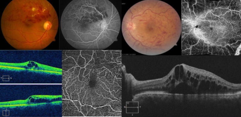
How is Retinal Vein Occlusion diagnosed?
Retinal Vein Occlusion can be easily detected by a trained ophthalmologist on dilated retinal examination. He/she may do the following tests to confirm the diagnosis.
- Every eye examination the following tests would be adviced by your doctor
- Visual aquity testing (distance and near)
- Detailed eye examination (slit lamp biomicroscopy)
- Dialated fundus examination (ophthalmoscopy / fundoscopy)
- Depending on your retinal findings, the doctor may advice for some special tests:
- Fundus photography
- Fundus Flurescein Angiography (FFA)
- Optical coherence tomography (OCT)
- OCT Angiography (OCT – A)
What are the treatment options for Retinal Vein Occlusion?
The treatment for Retinal Vein Occlusion is in the form of systemic control of general conditions (diabetes, cholesterol, and hypertension), and ocular treatments. The ocular treatment for Retinal Vein Occlusion is mainly for treatment of its complications (macular edema and retinal neo-vascularization) and is not meant for reversal or improvement of vision. The treatment options available are:
- Intravitreal Injections (Anti-VEGF, Steroids)
- Laser Treatment
- Surgery (for vitreous hemorrhage, epiretinal membrane)
Frequently Asked Questions (FAQ)
Yes. A blocked vein in the retina can cause many problems in the eye like:
- Macular edema:
When the vein is blocked, it may leak fluid in the retina. The center of the retina is called the macula. If blood leaks around the macula, it can cause swelling. This causes blurred vision or vision loss.
- Neovascularization:
To make up for the blocked veins, the eye may start to grow new, abnormal blood vessels. These may also leak fluid into the eye. In some cases, the new vessels may also pull on the retina and separate it from the back of the eye.
- Glaucoma:
When new blood vessels grow in certain areas of the eye (iris), they increase the eye pressure leading to glaucoma. Too much pressure can damage the optic nerve.
This varies from person to person. Over time, about 2/3rd of patients will regain at least some vision (1/3rd will rapidly improve, while the other 1/3rd will improve more gradually). For the best outcomes, all patients should follow their treatment plan. Follow-up visits with your eye doctor should be made as informed by your treating doctors. Your general physician should also be informed of any blood clots in the eyes.