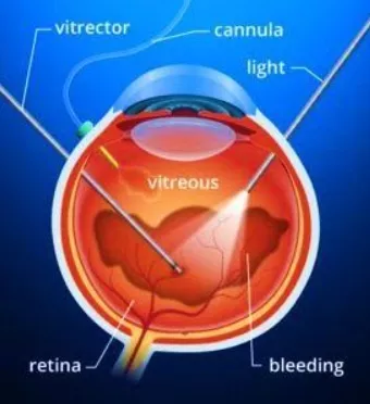
Vitreo - Retinal Surgery
What is Vitrectomy Surgery ?
The vitreous is a clear, jelly-like fluid (vitreous) that fills the back of the eye and gives shape to the eyeball just like air gives shape to a balloon. But in some diseases, it becomes cloudy or starts pulling on the retina. In these cases, the vitreous may need to be replaced. Vitrectomy is the surgery used to remove and replace this fluid and treat certain retinal diseases.

What are the conditions treated with Vitrectomy Surgery?
- Vitreous haemorrhage
- Severe eye trauma
- Diabetic Retinopathy
- Retinal Detachment
- Retinopathy of Prematurity
- Epiretinal Membrane
- Macular Hole
How is the operation performed?
The surgery may be done under general anaesthesia (sound asleep) or under local anaesthesia (you are awake but feel no pain). The operation usually takes 1-2 hrs. Once the vitreous is removed from the eye and the retinal problem taken care of, the vitreous is immediately replaced by salt solution, gas, or silicone oil. Tiny stitches may be used to close the wound. Most patients do not need to stay in hospital after surgery. A face-down position for sleeping may be suggested for several days. The operated eye will be bandaged for one day. Occasionally, both eyes may need to be bandaged to ensure complete ocular rest.
What is expected from me after the Surgery?
After surgery, the salt solution or gas bubble will slowly disappear. It will be replaced by fluid made by the eye. If your eye was filled with silicone oil (instead of salt solution or gas), the oil must be removed in 3-6 months. For the first two weeks you should rest at home. Travelling should be avoided except to see the doctor. If gas has been injected into the eye, you should avoid air travel for several weeks until specifically authorized by the doctor. Postoperative instructions will be given to you at the time of discharge and these should be strictly followed. Most patients are able to return to their routine in four weeks.
What are the chances of success?
The vision improves to some degree in 90% of simple vitrectomy cases. In difficult cases however, improvement is seen in approximately 60% of the cases while in others it may remain the same or even decrease. The final degree of clarity of vision is usually not evident for about three months. How much vision a patient will ultimately have is difficult to predict in individual cases. Patients are usually able to see large objects but fine vision and reading vision may not improve. Usually one operation is sufficient, occasionally additional surgery may be required.
What are the side effects and possible complications of surgery?
Blurred vision, pain in the eye, redness and swelling, mucus discharge and enlarged pupil and double vision are usually the temporary side effects and they clear up in a few weeks. Usually there are no complications, but some patients may have problems such as recurrent bleeding, infection, or elevated pressure in the eye. Rarely, a retinal detachment or cataract may develop requiring further surgery, either during or after the vitrectomy operation. Very rarely a complication may lead to the loss of all vision. To find out about anesthesia related complications, one may consult and anesthetist.
What are the precautions to be observed after Vitrectomy?
The following precautions should be observed for three weeks:
- Do not lift anything that weighs more than five kilograms.
- Do not bend over so that your head is below your waist.
- Avoid sleeping on the operated side, unless you are instructed to do so.
- Avoid sexual intercourse.
- Do not rub the operated eye.
- Avoid vigorous activity.
- No automobile trips except to visit the doctor.
- You may bathe carefully from below your neck and shave, but do not get the eyeball wet for at least two weeks. You may carefully clean the forehead and cheek with a wet cloth.
- You may watch television sparingly.
- You may gently rub the eyelids with cotton or a clean tissue moistened with warm water.
How do I maintain position after Vitrectomy Surgery?
- Certain retinal detachments require the placement of a bubble of sterile gas in the interior of the eyeball. When you observe a face-down position this gas rises toward the ceiling and pushes the retina back into place. The gas will be absorbed by the eye within a few weeks.
- You will be asked to lie on your side while in the recovery room.
- After moving to your ward bed, you will be placed in (A) Place a pillow under the chest and a rolled-up towel (or small blanket) under the forehead. (B) Lie on your side with your face turned toward the mattress. NOTE: The long axis of the head should always be parallel to the floor.
- You may lie on your back for 3 – 4 minutes for changing of the eye bandage, putting eye drops or for the doctor’s examination.
- For the first 10 days after surgery, you have a choice of five positions:
- Prone
- On side
- Sitting with elbows on knees
- Sitting with forehead on table edge, with small pillow
- Walking in the house with the long axis of head parallel to the floor
- Eat facing down, drink beverages with a straw

Scleral Buckling
What is Scleral Buckling surgery?
The surgery may be done under general anaesthesia (you will be sound asleep) or local anaesthesia (you will be awake but an injection will prevent any pain). The retina is reattached by freezing (cryosurgery) and with the placement of a permanent silicon patch (buckle) on the wall of your eyeball. The external stitches will melt away and do not have to be removed. Usually the eye responds to one operation; occasionally, additional surgery may be required. Normally, only the operated eye is bandaged but, sometimes, both eyes may be bandaged for a few days. Most patients can return to work in four to five weeks.
What may I do after surgery?
You must stay at home for at least three weeks, traveling should be avoided except to visit the doctor. After surgery you will be given written instructions regarding medication and precautions to be taken. You should carefully observe these instructions. You may be advised to lie on your side or stomach while sleeping or resting.
What are the chances of success?
In most cases (85%) the retina can be reattached with a single operation. Occasionally, additional surgery is necessary; this brings the final cure rate up to approximately 95%. The final degree of clarity of vision will not be known for three months. If you had lost your reading vision before surgery, you should find considerable improvement but probably not 100%. If your reading vision was not lost before surgery, good vision will be retained (after convalescence) in more than 90% cases. In 5% cases the retina may not re-attach, necessitating further surgery.
What are the common side effects and complications of the surgery?
Your vision will be blurred. The eye will be painful, red and swollen and there may be some mucus discharge. The pupil will be dilated and you may see double. These side effects are usually temporary and last only a few weeks. In many cases the eye will become more near-sighted; this can be corrected with spectacles. Over 90% cases have no significant complications. Occasional problems include bleeding or infection or re-detachment. Very rarely such complications could lead to the loss of all vision. Anaesthesia-related complications are also rare; the anesthetist will discuss these with you.
What about the future of my retina?
If the retina remains attached for three months after surgery, the chance of recurrence of retinal detachment is only 10%. If the retina of your other eye appears normal at this time, the chance of developing a detachment later on is approximately 12% in the eye that has not been operated.
Frequently Asked Questions (FAQ)
If I get vitrectomy surgery to fix a retinal detachment, will I regain my vision fully?
- It depends. If the centre of the retina was damaged, or if the detachment was not treated in early stages, complete visual recovery cannot be guaranteed. But, there is a chance that your vision will gradually improve.
Will I have any side effects from the silicon oil in my eye?
- A few patients might experience double vision. This improves after the oil is removed (3-6 months after surgery).
What will happen if the silicon oil is left in the eye?
- There is a high chance that cataracts will form. The eye pressure can also increase, causing nerve damage and eye pain.
Can I have a gas injection? I don’t want silicon oil because I have to get another surgery to remove it.
- That decision can only be made by a surgery. Based on the condition of your eye, one option might be better than the other. It is better to use the filling that is best for your eye health than to try and avoid another surgery.
What will happen if I choose to not get the surgery?
- The retinal condition will progress. You may lose your vision completely as the retina becomes more damaged. You can also develop cataract. If you have uveitis or glaucoma, you may experience eye pain. In the long run, your eyes might even shrink. Surgery offers a chance to improve your sight. Without it, your eyes will only get worse.
How many days will I have to tilt my head and lie on my stomach?
- For 7-15 days.

Contact
Address: 20, Farmland, Central Bazar Road, Near Lokmat Square, New Ramdaspeth, Nagpur – 440010, Maharashtra, India. Phone:+91 712 2425 839/2425 869/2425 899 For Appointment:+91 78755 20005 WhatsApp:+91 78755 10002 Email:anjanieyehospital1976@gmail.com Hospital Working Hours: 7:30 AM – 5:30 PM (Monday to Friday) 7:30 AM – 3:30 PM (Saturday) Sunday Closed
Copyright © 2025. All rights reserved. Built by SHOUT IN & OUT
Privacy Policy
